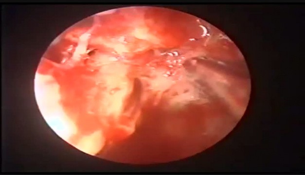
Cholesteatoma Of Attic Definition
Https Amc Edu Academic Gme Programs Otolaryngology Upload 2 Cholesteatoma Pdf

Cholesteatoma Long Case Ent Osce Dnb Mentors

Eardrums Seen In 8 Conditions Normal Eardrum Acute Otitis Media Perforation Small Perforation Attic Perforat Otitis Otitis Media Health Assessment Nursing
Cholesteatoma
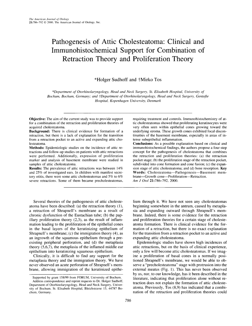
Recurrent Cholesteatoma In The Attic Arrow Download Scientific Diagram

Cholesteatoma Ento Key
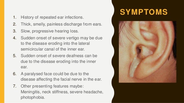
If untreated a cholesteatoma can eat into the three small bones located in the middle ear the malleus incus and stapes collectively called ossicles which can result in nerve deterioration.
Cholesteatoma of attic definition.
Chronic Otitis Media Causes Complications Management Teachmesurgery
Csom Cholesteatoma
Recidivism Ento Key

Pdf Cholesteatoma Definition And Classification A Literature Review Semantic Scholar

Photographs Retracted Eardrums Retraction Pockets Cholesteatomas Eardrum Perforations Serous And Acute Otitis Media Ear Fluid
Pdf Pathogenesis Of Attic Cholesteatoma Clinical And Immunohistochemical Support For Combination Of Retraction Theory And Proliferation Theory
General Considerations In Cholesteatoma Ento Key

Tympanic Membrane Retraction Classification Note Sade Grade 3 Retracted Tympanic Membrane Touching Promontory Toss Grad Membrane Sade Classification

Otitis Media Chronic Suppurative Ear Nose And Throat Disorders Merck Manuals Professional Edition
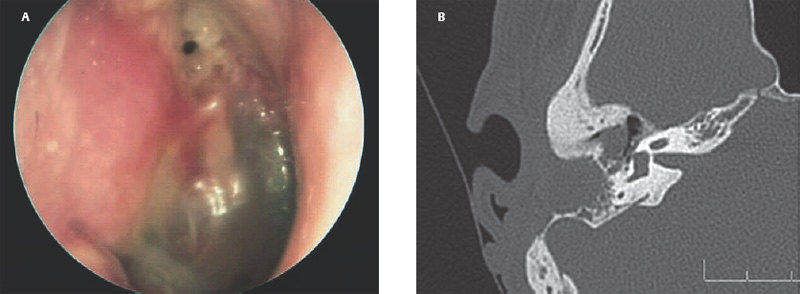
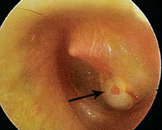
Attic Cholesteatoma With Csf Otorrhoea Video Medtube Net
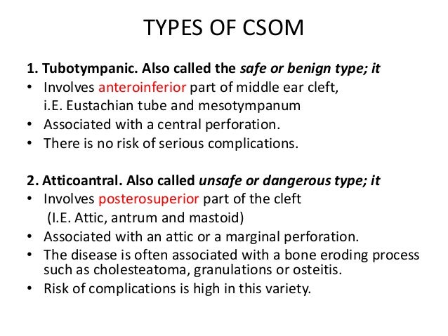
15 Chronic Suppurative Otitis Media Attico Antral Disease

Pdf Transcanal Endoscopic Mastoid Surgery With Tympanoplasty For The Management Of Cholesteatoma And Its Related Lesions Of Mastoid Antrum

Medical Tips And Info Otitis Otitis Media Health Assessment Nursing
Cholesteatoma Cme
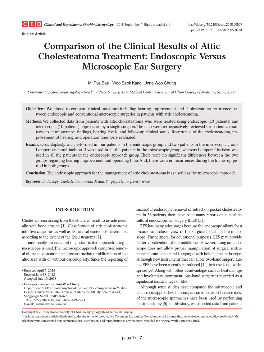
Pdf Comparison Of The Clinical Results Of Attic Cholesteatoma Treatment Endoscopic Versus Microscopic Ear Surgery

Figure 1 From Implementation Of The Eaono Jos Definitions And Classification Of Middle Ear Cholesteatoma From Stam To Stamco Semantic Scholar

Medpix Case Primary Cholesteatoma
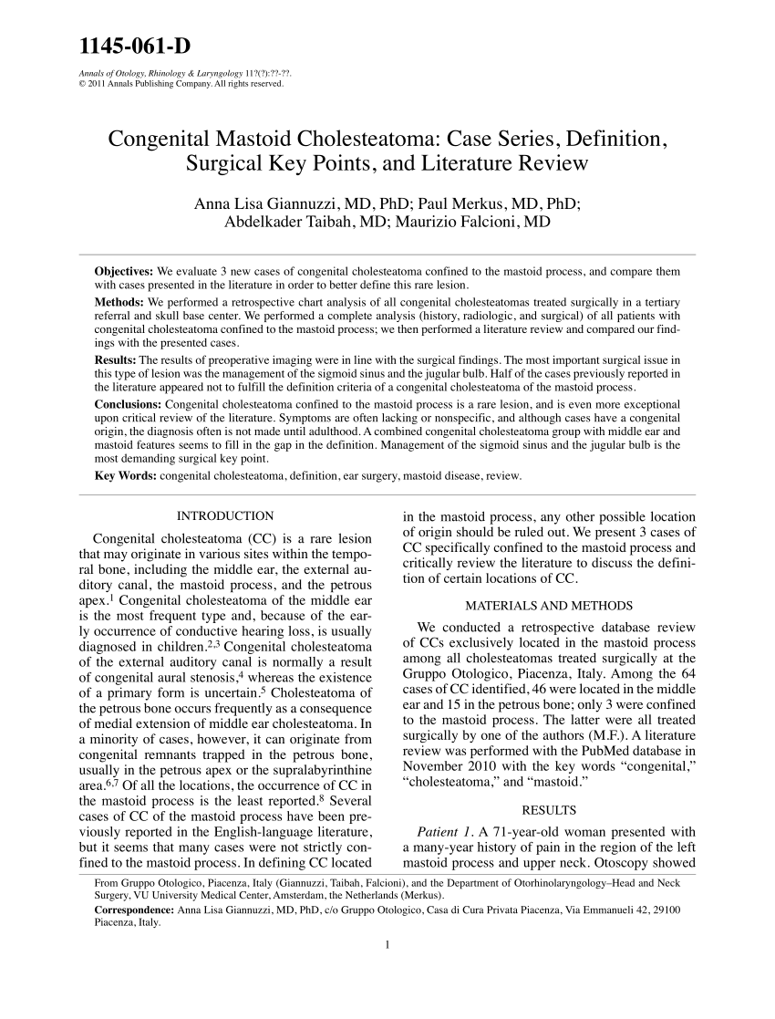
Pdf Congenital Mastoid Cholesteatoma Case Series Definition Surgical Key Points And Literature Review
Https Encrypted Tbn0 Gstatic Com Images Q Tbn 3aand9gcshxigp1cp Lksw5eyrjfubeo7r11ccyg1sj86hrxlmpnhx Awv Usqp Cau

Cao Staging System Of Cholesteatoma Download Table
Gale Academic Onefile Document Correlation Between Surgical Outcome And Stage Of Acquired Middle Ear Cholesteatoma Revalidation Of The Eaono Jos Staging System

Molecular Biological Diagnosis Of Congenital And Acquired Cholesteatoma On The Basis Of Differences In Telomere Length Kojima 2001 The Laryngoscope Wiley Online Library

Pdf Updates And Knowledge Gaps In Cholesteatoma Research
Https Chole Surgery Wp Content Uploads 2018 11 Introducing The Chole Classification Linder Pdf
Source : pinterest.com
